

Home > Company News
Gallstones are a common disease, often discovered during physical examinations. They may not cause any physical symptoms, or they may present with symptoms that go unnoticed. But once gallstones are discovered, is surgery always necessary? When is surgical treatment for gallstones required?
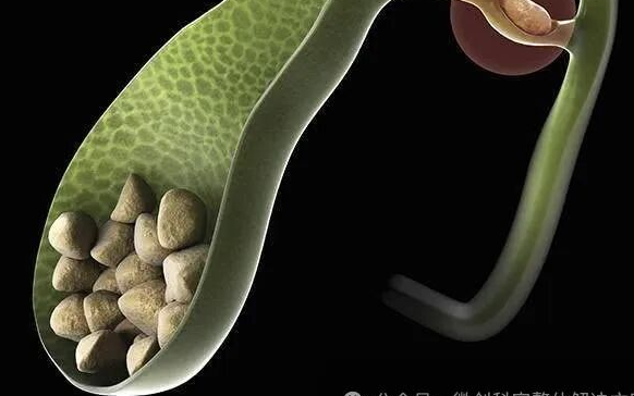
With the improvement of people's living standards and changes in dietary structure, the incidence of gallstones has increased. Gallstones are divided into gallbladder stones and bile duct stones. Bile duct stones are further divided into intrahepatic bile duct stones and extrahepatic bile duct stones. Extrahepatic bile duct stones are often accompanied by gallbladder stones. So, what kind of gallstones require surgical treatment?
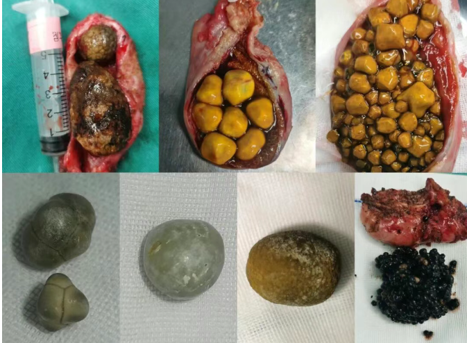
Simple gallbladder stones without any symptoms are mostly discovered during physical examinations and are called "quiet stones." Quiet stones generally do not require treatment. However, some argue that quiet stones should also be treated as early as possible because long-term stone irritation can induce gallbladder cancer.
Surgery is recommended for gallbladder stones complicated by acute or chronic inflammation, large stones, gallbladder polyps, or complications such as biliary pancreatitis. According to guidelines, laparoscopic cholecystectomy is the preferred treatment for gallstones. For patients who cannot tolerate surgery, gallbladder puncture and drainage can be considered.
For patients with gallstones complicated by common bile duct stones, early treatment is strongly recommended. Treatment methods mainly include laparoscopic cholecystectomy with ERCP stone removal and laparoscopic cholecystectomy combined with choledochoscopy stone removal. Each method has its advantages and disadvantages, and an individualized treatment plan should be developed based on the patient's specific condition. Specific steps of gallstone surgery:
STEP 1: Make the first incision at the umbilicus, insert a trocar, and place a camera.
STEP 2: Make the remaining three incisions in the upper abdomen and insert trocars.
STEP 3: Locate the gallbladder and grasp it with clamps.
STEP 4: Dissect the fat to expose two important structures—the cystic duct and the cystic artery.
STEP 5: Clamp and cut the cystic duct and cystic artery.
STEP 6: Begin dissecting the gallbladder from behind the liver.
STEP 7: Perform the procedure gently until the gallbladder is completely removed.
STEP 8: Place the gallbladder in this small bag and remove it through the incision in the upper abdomen.
STEP 9: Gallstones.
Key Points and Challenges of Laparoscopic Cholecystectomy
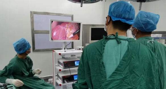
Good surgical visualization is a prerequisite for proper handling of the structures within Calot's triangle. Proper handling of the structures within Calot's triangle is crucial for successful cholecystectomy, and the quality of this handling directly affects the patient's prognosis.
1. Good Exposure of Calot's Triangle
① After inserting the laparoscope to understand the liver's position, make a trocar incision on the right side of the falciform ligament below the xiphoid process, perpendicular to or slightly below the lower edge of the liver. The distance between the xiphoid trocar incision and the trocar incision at the right midclavicular line should be approximately 10 cm. The trocar incisions at the midclavicular line and the costal margin below the axillary line should be slightly below the lower edge of the liver.
② Changes in body position and pneumoperitoneum pressure: For patients with abundant intra-abdominal fat, poor bowel preparation, and gastrointestinal distension, the greater omentum and gastrointestinal tract shift upwards, narrowing the subhepatic space and resulting in poor exposure of the Calot's triangle. In such cases, the intra-abdominal pressure can be appropriately increased to 15 mmHg. During the operation, the patient should be positioned with their head elevated and feet lowered, tilted 15 degrees to the left. The gravity of the aforementioned organs and adipose tissue helps to widen the subhepatic space and increase the operating space.
2. Dissection and Management of Tissues within the Calot's Triangle
The Calot's triangle should be kept as open as possible. The area around the confluence of the gallbladder ampulla and cystic duct should be fully dissected to clearly display the internal structures. The following problems may be encountered during the operation:
① If adhesions in the Calot's triangle are severe, the following points should be noted during dissection: Adhesion dissection should be performed under tension traction close to the gallbladder ampulla, with gentle manipulation. Blunt dissection methods (such as dissecting forceps, non-electrostatic hooks, and miniature electric shears) are generally used to avoid blind electrocoagulation and electrocautery. Cases of extrahepatic bile duct burns due to heat have been frequently reported.
In cases of acute cholecystitis (or with impacted gallstones), Calot's triangle often presents with significant congestion and edema, but adhesions are not severe. During the dissection of the gallbladder ampulla and Calot's triangle, extravasation of edema fluid is common. A blunt dissection with the tip of an irrigator can be used while repeatedly irrigating to maintain a clear view, increasing the likelihood of surgical success.
When there are seamless adhesions between the common bile duct, common hepatic duct, right hepatic duct, and gallbladder ampulla, the serosa can be incised at the junction of the gallbladder ampulla and the cystic duct. Dissection forceps can be used above the cystic duct to expose Calot's triangle. If the cystic duct still cannot be exposed, retrograde cholecystectomy can be performed.
② The cystic duct exhibits considerable anatomical variation in thickness and length. When exposing and managing it, the following principles should be followed: The area around the junction of the cystic duct and the ampulla of Vater must be fully dissected. If the "three ducts and one ampulla" (common hepatic duct, common bile duct, cystic duct, and ampulla of Vater) cannot be fully visualized, there should be an empty space above the confluence of the cystic duct and the common hepatic duct (meaning the common hepatic duct is not within this space) when clamping the cystic duct.
After clamping, the cystic duct should be cut to avoid burning the extrahepatic bile ducts due to heat. If the cystic duct is significantly thickened and direct management is difficult, retrograde gallbladder resection and ligation with a snare can be performed.
③ The course and branches of the cystic artery exhibit many anatomical variations. It is crucial to avoid treating one branch while neglecting others. It is best to avoid "skeletalizing" the cystic artery to prevent insufficient vascular tissue and insecure clamping. When encountering large vascular branches during dissection of the gallbladder bed, clamping should be applied for hemostasis, especially when there is no main cystic artery trunk within Calot's triangle. Note the presence of an artery behind the cystic duct. If a noticeable resilient feeling is felt when separating the posterior part of the cystic duct, this condition should be suspected. In this case, the upper half of the clamped cystic duct can be cut off first, and another clamp can be placed on the distal side of the cut cystic duct. Then, the entire cystic duct (including the cystic artery behind the cystic duct) can be cut off.

+86-13167065777
Building 5A, No. 9628, Hunan Highway, Pudong New Area, Shanghai, China
 Links:
Shanghai Shiyin Photoelectric Instrument Co.,Ltd. All Right Reserved