

Home > Company News
Epistaxis is a frequent clinical issue in otolaryngology and a secondary symptom of various diseases, accompanied by complicated causative factors. As a common emergency in the ENT department, its core treatment target is to quickly find bleeding spots and implement effective hemostasis measures. Medical history records show that nasal bleeding treatments have existed for over a thousand years; traditional therapy mainly depends on nasal packing to stop hemorrhage. Nevertheless, this conventional measure brings great pain to patients, often fails to reach ideal hemostatic effects, and may trigger multiple complications, especially for patients suffering from hypertension and coronary heart disease. For stubborn and recurring severe epistaxis, common treatment choices include anterior and posterior nasal packing embolization, arterial ligation and vascular embolization therapy. Digital subtraction angiography (DSA) is a minimally invasive technology combining diagnosis and treatment with promising clinical prospects, which works best for epistaxis caused by arterial malformations and nasal aneurysms. After locating lesions via angiography, embolization can effectively stop bleeding. However, DSA barely works for epistaxis triggered by other causes. Besides, its operation steps are complicated and carry relatively high risks, so clinicians must follow strict standards when selecting DSA for patients.
Kiesselbach's area of the nasal septum
Wu's venous plexus at the posterior inferior nasal meatus
Posteroinferior part of the nasal septum
Anterior wall of the sphenoid sinus (edge of posterior nasal aperture)
Nasal roof (olfactory cleft)
Other hidden areas
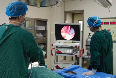
Endoscopic micro-packing: Relying on the clear sight, sufficient light and precise positioning of nasal endoscopy, medical staff perform targeted micro-packing with petrolatum gauze, hemostatic gauze or hemostatic fiber after confirming bleeding foci. This method boasts high hemostasis efficiency, retains normal nasal ventilation, greatly reduces patients’ discomfort, and avoids redundant posterior nasal packing for posterior nasal bleeding.
Endoscopic high-frequency electrocoagulation: After pinpointing bleeding positions (especially broken small vascular stumps), the high-frequency electrode generates electric arcs to form local high temperature, carbonizing tissues and sealing broken blood vessels to achieve hemostasis.
Laser-assisted endoscopic hemostasis: Endoscopic laser carbonization and vascular closure are widely applied in clinical epistaxis treatment. Common laser devices include Nd:YAG laser, CO2 laser, KTP532 laser, semiconductor laser and holmium laser, among which Nd:YAG laser and KTP532 laser are most widely used in clinical practice.
Endoscopic microwave coagulation hemostasis: Microwaves belong to high-frequency electromagnetic waves, which can instantly raise local tissue temperature, lead to tissue denaturation and coagulation, and rapidly stop bleeding. The microwave probe can directly contact bleeding spots for quick hemostasis.
Low-temperature plasma radiofrequency ablation hemostasis: This technology follows the low-temperature coagulation principle. It uses plasma radiofrequency energy at 4–70°C to coagulate bleeding points and stop hemorrhage. Operators should control coagulation time properly and stop the operation once the nasal mucosa turns white or faint yellow. When treating nasal septum bleeding with this method, great attention should be paid to prevent the complication of nasal septum perforation.
It can accurately find active bleeding points in all parts of the nasal cavity, especially hard-to-detect lesions in the posterior nasal cavity.
It supports precise, intuitive and simple operations under direct vision, realizing fast, accurate and effective hemostasis with reliable curative effects.
It causes minimal tissue damage and pain for patients, avoids unnecessary anterior and posterior nasal packing, so it is especially suitable for epistaxis patients with hypertension, vascular diseases and blood system disorders.

+86-13167065777
Building 5A, No. 9628, Hunan Highway, Pudong New Area, Shanghai, China
 Links:
Shanghai Shiyin Photoelectric Instrument Co.,Ltd. All Right Reserved